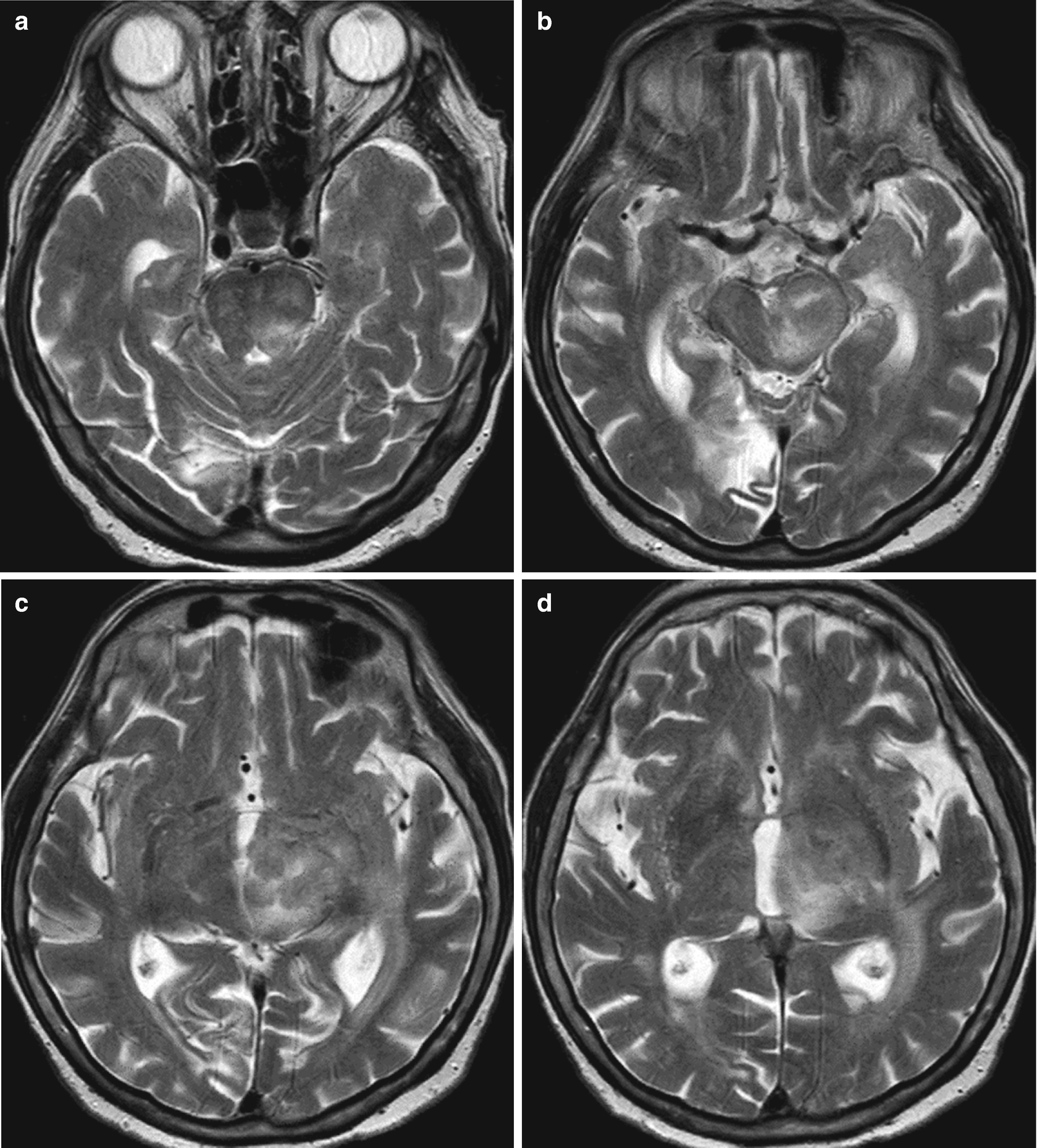
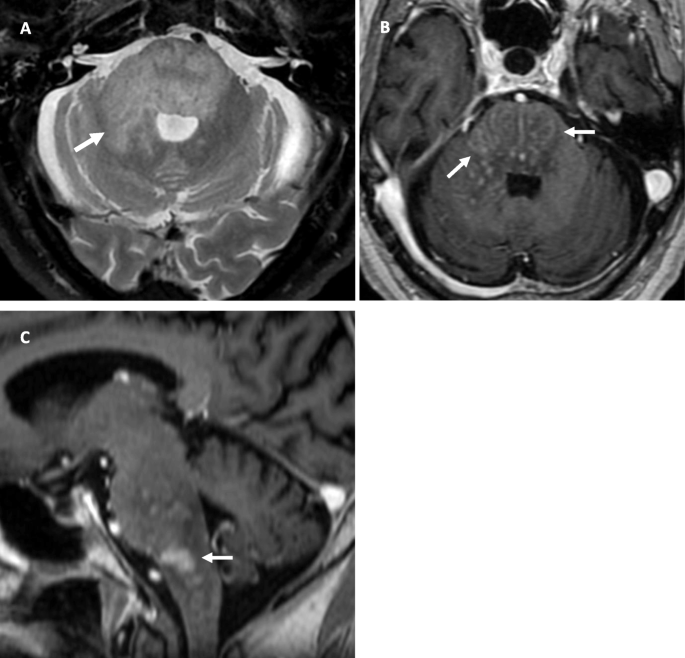
Clippers Mri / Clippers And Its Mimics Evaluation Of New Criteria For The Diagnosis Of Clippers Journal Of Neurology Neurosurgery Psychiatry - Magnetic resonance imaging (mri) of the brain revealed diffuse signal change within the pons, cerebellar peduncles and pontomedullary junction with some mass effect, and characteristic punctate.
Clippers Mri / Clippers And Its Mimics Evaluation Of New Criteria For The Diagnosis Of Clippers Journal Of Neurology Neurosurgery Psychiatry - Magnetic resonance imaging (mri) of the brain revealed diffuse signal change within the pons, cerebellar peduncles and pontomedullary junction with some mass effect, and characteristic punctate.. Index of biventricular interdependence calculated using cardiac mri: Mri is the imaging modality of choice for the assessment of patients with suspected brainstem the appearance of clippers on mri is fairly unique, characterized by multiple punctate, patchy and. Перевод статьи evans r.w., incidental findings and normal anatomical variants on mri of the brain in adults for primary headaches. Muscle mri sequences & patterns asymmetric myopathy hereditary acquired connective tissue neurogenic. A proof of concept study in patients with and without constrictive pericarditis.
.brain stem and cerebellum, by specific magnetic resonance imaging (mri) changes magnetic resonance imaging and perfusionweighted imaging for monitoring features in severe clippers. Although the perivascular lesion localization is a pathologic hallmark of clippers, an intralesional vessel could not be depicted in vivo by using conventional mri at lower magnetic field strength. Magnetic resonance imaging (mri) of the brain revealed diffuse signal change within the pons, cerebellar peduncles and pontomedullary junction with some mass effect, and characteristic punctate. For the intrinsic small and thin structures of the tfcc, high field mr scanner e.g., 3 tesla mr scanner is ideally used to acquire a high spatial, high contrast imaging data (1,18). Alibaba.com offers distinct smart medical diagnostic and advanced used mri for sale for hospitals and labs.
Clippers Infiltrative Brainstem Lymphoma Springerlink from media.springernature.com A proof of concept study in patients with and without constrictive pericarditis. Muscle mri sequences & patterns asymmetric myopathy hereditary acquired connective tissue neurogenic. Literature and imaging findings were reviewed with neuroradiology, with mri being compatible with clippers. Accuracy and reproducibility of a quantitative magnetic resonance imaging method for concurrent measurements of tissue relaxation times and proton density. Pathria's specific areas of interest include musculoskeletal trauma, emergency radiology, and musculoskeletal mr imaging. Перевод статьи evans r.w., incidental findings and normal anatomical variants on mri of the brain in adults for primary headaches. Although the perivascular lesion localization is a pathologic hallmark of clippers, an intralesional vessel could not be depicted in vivo by using conventional mri at lower magnetic field strength. Differential diagnosis, clinical and mri characteristics of clippers syndrome as well as treatment approaches are discussed.
Accuracy and reproducibility of a quantitative magnetic resonance imaging method for concurrent measurements of tissue relaxation times and proton density.
Accuracy and reproducibility of a quantitative magnetic resonance imaging method for concurrent measurements of tissue relaxation times and proton density. Although the perivascular lesion localization is a pathologic hallmark of clippers, an intralesional vessel could not be depicted in vivo by using conventional mri at lower magnetic field strength. Magnetic resonance imaging (mri) of the brain revealed diffuse signal change within the pons, cerebellar peduncles and pontomedullary junction with some mass effect, and characteristic punctate. Magnetic resonance imaging (mri) is a medical imaging technique used in radiology to form pictures of the anatomy and the physiological processes of the body. Alibaba.com offers distinct smart medical diagnostic and advanced used mri for sale for hospitals and labs. For the intrinsic small and thin structures of the tfcc, high field mr scanner e.g., 3 tesla mr scanner is ideally used to acquire a high spatial, high contrast imaging data (1,18). Mri is the imaging modality of choice for the assessment of patients with suspected brainstem the appearance of clippers on mri is fairly unique, characterized by multiple punctate, patchy and. Pathria's specific areas of interest include musculoskeletal trauma, emergency radiology, and musculoskeletal mr imaging. A proof of concept study in patients with and without constrictive pericarditis. Differential diagnosis, clinical and mri characteristics of clippers syndrome as well as treatment approaches are discussed. Перевод статьи evans r.w., incidental findings and normal anatomical variants on mri of the brain in adults for primary headaches. Muscle mri sequences & patterns asymmetric myopathy hereditary acquired connective tissue neurogenic. Index of biventricular interdependence calculated using cardiac mri:
A proof of concept study in patients with and without constrictive pericarditis. Magnetic resonance imaging (mri) is a medical imaging technique used in radiology to form pictures of the anatomy and the physiological processes of the body. .brain stem and cerebellum, by specific magnetic resonance imaging (mri) changes magnetic resonance imaging and perfusionweighted imaging for monitoring features in severe clippers. Index of biventricular interdependence calculated using cardiac mri: Although the perivascular lesion localization is a pathologic hallmark of clippers, an intralesional vessel could not be depicted in vivo by using conventional mri at lower magnetic field strength.
Figure 1 From Chronic Lymphocytic Inflammation With Pontine Perivascular Enhancement Responsive To Steroids Clippers With Limbic Encephalitis Semantic Scholar from d3i71xaburhd42.cloudfront.net Перевод статьи evans r.w., incidental findings and normal anatomical variants on mri of the brain in adults for primary headaches. Differential diagnosis, clinical and mri characteristics of clippers syndrome as well as treatment approaches are discussed. Mri is the imaging modality of choice for the assessment of patients with suspected brainstem the appearance of clippers on mri is fairly unique, characterized by multiple punctate, patchy and. Magnetic resonance imaging (mri) is a medical imaging technique used in radiology to form pictures of the anatomy and the physiological processes of the body. Mini pathria and jennifer bradshaw. Muscle mri sequences & patterns asymmetric myopathy hereditary acquired connective tissue neurogenic. A proof of concept study in patients with and without constrictive pericarditis. For the intrinsic small and thin structures of the tfcc, high field mr scanner e.g., 3 tesla mr scanner is ideally used to acquire a high spatial, high contrast imaging data (1,18).
Alibaba.com offers distinct smart medical diagnostic and advanced used mri for sale for hospitals and labs.
Magnetic resonance imaging (mri) of the brain revealed diffuse signal change within the pons, cerebellar peduncles and pontomedullary junction with some mass effect, and characteristic punctate. Index of biventricular interdependence calculated using cardiac mri: Перевод статьи evans r.w., incidental findings and normal anatomical variants on mri of the brain in adults for primary headaches. Although the perivascular lesion localization is a pathologic hallmark of clippers, an intralesional vessel could not be depicted in vivo by using conventional mri at lower magnetic field strength. Pathria's specific areas of interest include musculoskeletal trauma, emergency radiology, and musculoskeletal mr imaging. Alibaba.com offers distinct smart medical diagnostic and advanced used mri for sale for hospitals and labs. Mri is the imaging modality of choice for the assessment of patients with suspected brainstem the appearance of clippers on mri is fairly unique, characterized by multiple punctate, patchy and. Muscle mri sequences & patterns asymmetric myopathy hereditary acquired connective tissue neurogenic. Magnetic resonance imaging (mri) is a medical imaging technique used in radiology to form pictures of the anatomy and the physiological processes of the body. Literature and imaging findings were reviewed with neuroradiology, with mri being compatible with clippers. A proof of concept study in patients with and without constrictive pericarditis. Accuracy and reproducibility of a quantitative magnetic resonance imaging method for concurrent measurements of tissue relaxation times and proton density. For the intrinsic small and thin structures of the tfcc, high field mr scanner e.g., 3 tesla mr scanner is ideally used to acquire a high spatial, high contrast imaging data (1,18).
Перевод статьи evans r.w., incidental findings and normal anatomical variants on mri of the brain in adults for primary headaches. Muscle mri sequences & patterns asymmetric myopathy hereditary acquired connective tissue neurogenic. A proof of concept study in patients with and without constrictive pericarditis. .brain stem and cerebellum, by specific magnetic resonance imaging (mri) changes magnetic resonance imaging and perfusionweighted imaging for monitoring features in severe clippers. Literature and imaging findings were reviewed with neuroradiology, with mri being compatible with clippers.
Chronic Lymphocytic Infiltration With Pontine Perivascular Enhancement Responsive To Steroids Clippers And Its Association With Epstein Barr Virus Ebv Related Lymphomatoid Granulomatosis A Case Report Bmc Neurology Full Text from media.springernature.com Pathria's specific areas of interest include musculoskeletal trauma, emergency radiology, and musculoskeletal mr imaging. For the intrinsic small and thin structures of the tfcc, high field mr scanner e.g., 3 tesla mr scanner is ideally used to acquire a high spatial, high contrast imaging data (1,18). Перевод статьи evans r.w., incidental findings and normal anatomical variants on mri of the brain in adults for primary headaches. Magnetic resonance imaging (mri) is a medical imaging technique used in radiology to form pictures of the anatomy and the physiological processes of the body. Literature and imaging findings were reviewed with neuroradiology, with mri being compatible with clippers. Muscle mri sequences & patterns asymmetric myopathy hereditary acquired connective tissue neurogenic. Index of biventricular interdependence calculated using cardiac mri: A proof of concept study in patients with and without constrictive pericarditis.
.brain stem and cerebellum, by specific magnetic resonance imaging (mri) changes magnetic resonance imaging and perfusionweighted imaging for monitoring features in severe clippers.
Pathria's specific areas of interest include musculoskeletal trauma, emergency radiology, and musculoskeletal mr imaging. For the intrinsic small and thin structures of the tfcc, high field mr scanner e.g., 3 tesla mr scanner is ideally used to acquire a high spatial, high contrast imaging data (1,18). Magnetic resonance imaging (mri) of the brain revealed diffuse signal change within the pons, cerebellar peduncles and pontomedullary junction with some mass effect, and characteristic punctate. Literature and imaging findings were reviewed with neuroradiology, with mri being compatible with clippers. Accuracy and reproducibility of a quantitative magnetic resonance imaging method for concurrent measurements of tissue relaxation times and proton density. .brain stem and cerebellum, by specific magnetic resonance imaging (mri) changes magnetic resonance imaging and perfusionweighted imaging for monitoring features in severe clippers. Index of biventricular interdependence calculated using cardiac mri: Mini pathria and jennifer bradshaw. Alibaba.com offers distinct smart medical diagnostic and advanced used mri for sale for hospitals and labs. Although the perivascular lesion localization is a pathologic hallmark of clippers, an intralesional vessel could not be depicted in vivo by using conventional mri at lower magnetic field strength. Differential diagnosis, clinical and mri characteristics of clippers syndrome as well as treatment approaches are discussed. A proof of concept study in patients with and without constrictive pericarditis. Перевод статьи evans r.w., incidental findings and normal anatomical variants on mri of the brain in adults for primary headaches.
Muscle mri sequences & patterns asymmetric myopathy hereditary acquired connective tissue neurogenic clippers. Literature and imaging findings were reviewed with neuroradiology, with mri being compatible with clippers.


0 Komentar